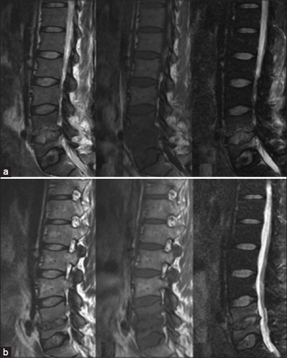
Figure 3.

Lumbar spine magnetic resonance imaging: (a) Fat-suppressed T2-weighted imaging (WI), T1-WI, and T2-WI showed pathological changes in L4 and L5 vertebrae and L4/L5 and L5/S1 intervertebral disks, showing vertebral osteomyelitis and diskitis (May 29, 2008). (b) Changes in lumbar vertebrae during the recovery period (October 13, 2008).
