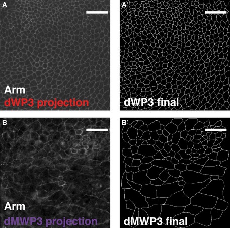
Figure EV5. Original images processed to obtain segmented data.

-
AProjection of the original confocal stack used to segment dWP3 sample.
-
A'dWP3 final segmented image.
-
BProjection of the original confocal stack used to segment dWP3 sample. Some confocal sections included signal from the peripodial membrane of the wing imaginal disc. Therefore, part of the processing was manual.
-
B'dMWP3 segmented final image. The signal from the peripodial membrane has been removed.
Data information: Scale bars: 10 μm.
