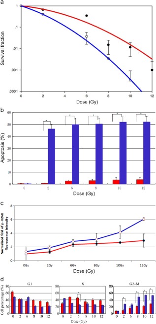
Figure 1.

Mutant epidermal growth factor receptor‐expressing cell lines show enhanced sensitivity to radiation. (a) Colonies were counted on the 14th day following radiation, and survival fractions were plotted as a function of dose. The survival curve was obtained by the linear‐quadratic model. Error bars, standard deviation (SD) from mean of triplicate measurements. (b) The apoptosis rate induced by radiation in H820 and A549 cells. Cells were stained with AnnexinV‐ fluorescein isothiocyanate/propium iodide (PI) at 36 hours following radiation; radiation‐induced apoptosis was higher in H820 than in A549 cells. (c) Cells were fixed at 24 hours after irradiation and probed with anti‐phosphorylated histone γ‐H2AX antibody to detect DNA repair capability. H820 cells show significantly more residual γ‐H2AX
, which indicates inefficient DNA repair. Normalized fold fluorescence intensity: the relative values of γ‐H2AX fluorescence intensity divided by unirradiated A549 cells. (d) A549 and H820 cells were stained with PI at 18 hours following radiation. Both cell lines revealed an increase of cells in G2/M with IR treatment. H820 cells show significantly more G2/M arrest and dismal S arrest compared with WT‐expressing cells. Error bars, SD from mean of triple independent measurements. (a) ( ) A549, (
 ) H820; (b) (
 ) A549, (
 ) H820; (c) (
 ) A549, (
 ) H820; (d) (
 ) A549, (
 ) H820.
