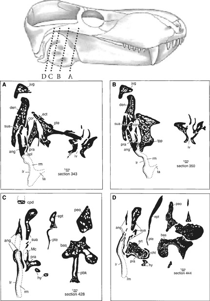
Figure 2.

Reconstructed recessus mandibularis (tympanic cavity) in primitive therocephalian therapsids. Above is the skull of Glanosuchus macrops, a scymnosaurine therocephalian (modified from Brink, 1988). Below are four cross‐sections of Glanosuchus sp. drawn from a grinding series housed at the Department of Zoology at the University of Stellenbosch: the section planes are indicated by the stippled lines in the figure of the skull of Glanosuchus as well as by letters A–D. The likely position of Meckel's cartilage is indicated by a dotted line, i.e. it was formed as cartilage; only its posterior end is ossified as articular. In the cross‐sections the hypothetical recessus mandibularis is drawn in by a stippled contour underneath the very thin bony plate of the reflected lamina of the angular. The proximal portions of the hyoids show the mammalian tympanohyal (from Maier & van den Heever, 2002).
