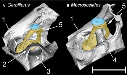
Figure 8.

MicroView reconstructions of the left stapes and associated structures of (A) Gerbillurus setzeri and (B) Macroscelides flavicaudatus, seen from a rostral, ventral and lateral position. The stapes is in each case shaded in yellow, the lenticular apophysis (part of the incus) in blue. Key: 1 = rim of oval window, containing the stapes footplate; 2 = bony collar surrounding course of stapedial artery; 3 = canal for stapedial artery; 4 = bony tube for stapedial artery; 5 = muscular process for the insertion of the m. stapedius on the stapes. Note that the enclosure of the stapedial artery within a bony tube is nearly complete in Macroscelides, but far less so in Gerbillurus. In Macroscelides, the stapes footplate fits the oval window more snugly than in Gerbillurus. Scale bar: 1 mm.
