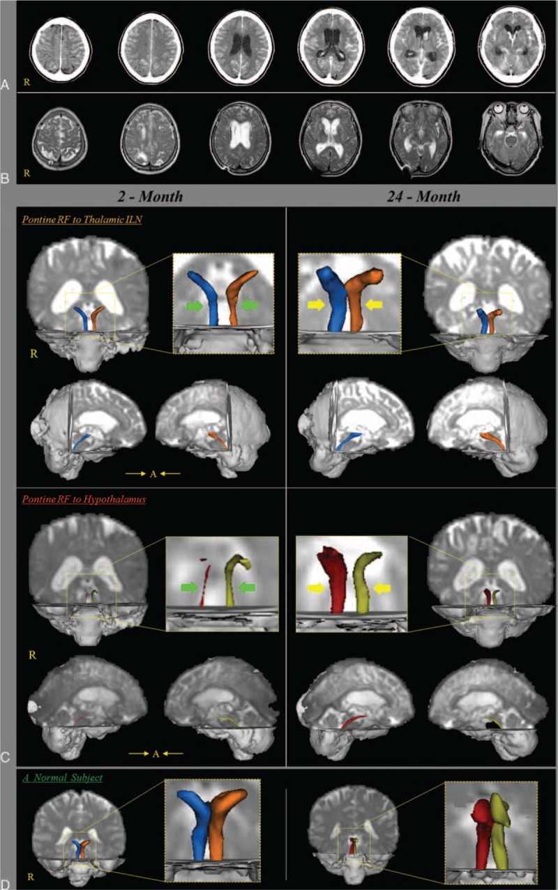
FIGURE 1.

(A) Brain CT images at onset show a subarachnoid hemorrhage and intraventricular hemorrhage and hydrocephalus. (B) Brain MR images at 2 months after onset show a leukomalactic lesion in both fronto-parietal lobes. (C) Results of diffusion tensor tractography (DTT) for both lower dorsal and ventral ascending reticular activating system (ARAS). On 2-month DTT, narrowing (arrows) of both lower dorsal and ventral ARASs was observed on both sides: in particular, among 4 neural tracts of the lower ARAS, the right lower ventral ARAS was narrowest. By contrast, on 24-month DTT, the 4 narrowed neural tracts of both lower dorsal and ventral ARASs were thickened compared with those of 2-month DTT (arrows). (D) DTTs of the lower dorsal and ventral lower ARAS of a normal subject (65-year-old woman). ARAS = ascending reticular activating system, CT = computed tomography, DTT = diffusion tensor tractography, MR = magnetic resonance.
