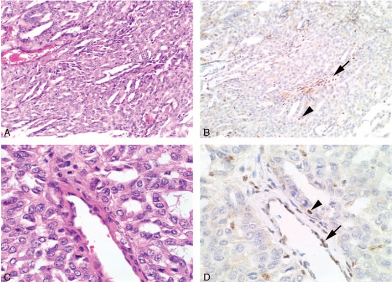
FIGURE 2.

Serial H&E (A, C) and BAP1 IHC (B, D) stained sections from an intrahepatic cholangiocarcinoma that shows completely negative IHC staining for BAP1. In this case, the nonneoplastic endothelial cells (arrows) and lymphocytes (arrowheads) demonstrated preserved positive staining and serve as an internal positive control (original magnifications A, B ×100, C, D ×400).
