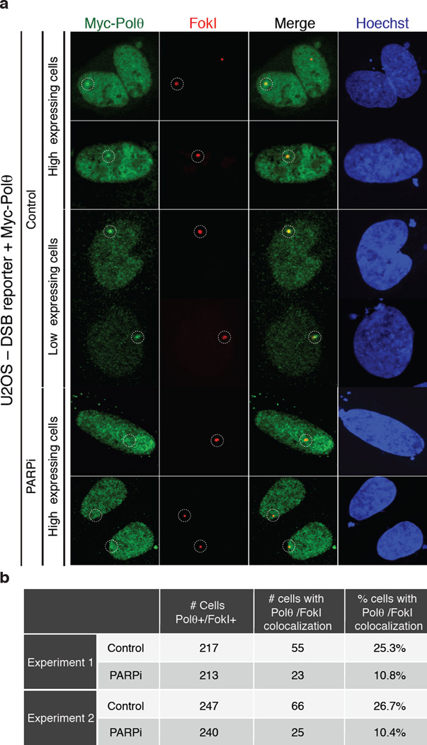
Extended data figure 7.
(Related to Figure 3). a, Results from immunofluorescence performed at 4 hours after induction (Shield1 ligand, Clontech 631037; 0.5 µM 4-OH tamoxifen) of DSBs by mCherry-LacI-FokI in the U2OS-DSB reporter cells18 transfected with the Myc-PolQ and treated with PARP inhibitor (KU58948). The mCherry signal is used to identify the area of damage and to assess the recruitment of Myc-PolQ to cleaved LacO repeats. b, Table displaying quantification related to a.

