Figure 1.
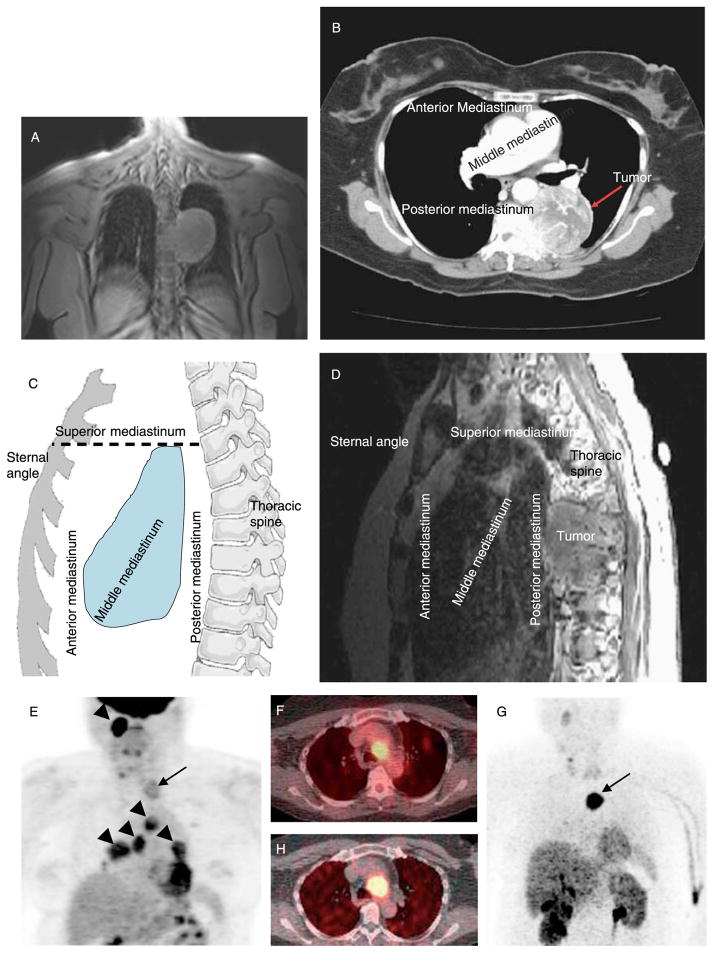
Imaging studies. (A) Chest MRI of patient 2 (coronal T1-weighted FOV three-plain localizing image; TR, 66.5 ms; TE, 1.5 ms) showing mediastinal mass. (B) Chest CT of patient 2 following i.v. contrast showing same paraganglioma. (C) Illustration of mediastinal compartments. The anterior mediastinum is defined as the space bordered by the sternum anteriorly and the ventral cardiac surface posteriorly, including the thymus and ascending aorta. The middle mediastinum is posterior to the anterior mediastinum and is bordered posteriorly by the anterior surface of the spine; the middle compartment includes the heart, esophagus, trachea, and major blood vessels. The posterior mediastinum is posterior to the anterior surface of the spine and contains the descending aorta, spine, and ribs. The superior mediastinum is defined by a horizontal line from the angle of Louis posteriorly to the spine as the inferior border and includes the thyroid, aortic arch, and superior parts of the esophagus and trachea. (D) MRI of patient 2 (sagittal T1-weighted images after gadolinium contrast; TR, 417 ms; TE, 13 ms) showing posterior mediastinal PGL. (E) Coronal [18F]FDG PET scan of patient 8, showing superior mediastinal PGL (arrow) plus additional lesions in the head, neck, and upper chest (arrowheads); axial CT-PET in (F) localizes mediastinal PGL near the aortic arch. (G) Coronal [18F]fluorodopamine PET scan of patient 8 shows localization of mediastinal PGL concordant with [18F]FDG PET (arrow), which was confirmed on axial CT-PET in (H).

