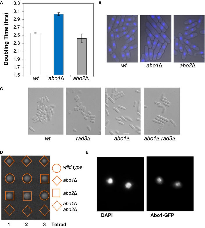
Figure EV1. Phenotypes associated with deletion of abo1 + .

- Doubling time was estimated by measuring the OD595 of the indicated strains grown in YE5S medium at 30°C. Data are the mean of two independent biological repeats, and error bars denote the range of the data.
- abo1Δ cells have an elongated morphology. Microscopic analysis of DAPI‐stained cells. Data are representative of three biological repeats.
- The elongated morphology of abo1Δ cells is independent of the ATR checkpoint kinase, Rad3. Microscopic analysis of the indicated strain. Images are representative of duplicate experiments.
- An abo1Δ abo2Δ double mutant strain is not viable. Tetrad dissection of a genetic cross between abo1Δ and abo2Δ. Colonies arising from the spores of three asci are shown. Data are representative of three biological repeats.
- Subcellular localisation of Abo1. Cells expressing Abo1‐GFP were stained with DAPI and visualised using fluorescence microscopy. Data are representative of two biological repeats.
