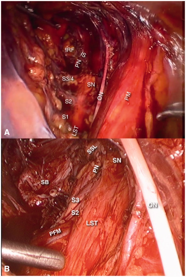
Fig. 3.
Nerves of the obturator space (right side). Picture (A) is the final aspect of a laparoscopic approach to Alcock’s Canal Syndrome, where the sacrospinous ligament has been transected to expose the pudendal nerve (PN). In picture B, the sacrospinous ligament (SSL) is intact. In both pictures, the internal and external iliac vessels are retracted medially. ON, obturator nerve; PM, psoas muscle; SN, sciatic nerve; LST, lumbosacral trunk; PN, pudendal nerve; IRF, ischiorectal fossa; IS, ischial spine; SB, sacral bone; PFM, pyriformis muscle.

