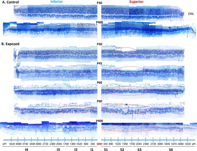
Fig 1. A representative reconstruction of the ONL of the superior (left) and inferior (right) retina (composed of 12–14 consecutive histological segments of 75μm in width, each sectioned at every 340μm from the ONH to the ora serrata for each hemiretina) obtained from control (A) and light exposed (B) juvenile rats at selected postnatal ages [from P30 to P400].
Abbreviations: ONL: outer nuclear layer; ONH: optic nerve head; S1 to S4: Sector 1 to 4 for the superior retina; I1 to I4; Sector 1 to 4 for the inferior retina. The extent of the retinal hole is highlighted with red dashed circles in the superior hemiretina.

