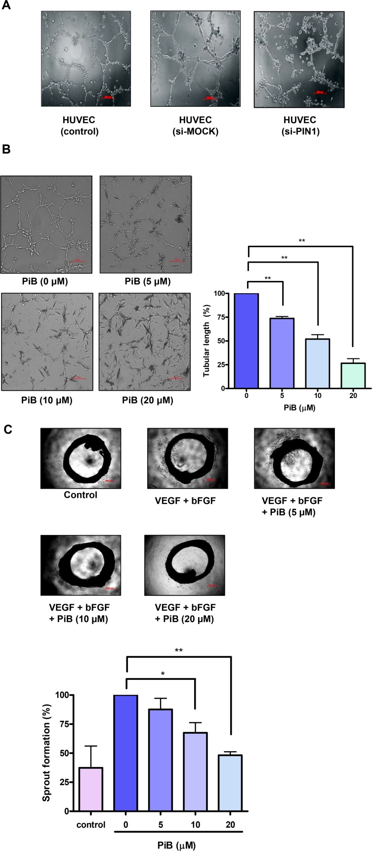
Fig 6. PIN1 enhanced 3D tube formation and ex vivo aortic ring vascular sprouting.
A) HUVEC were transfected with control or PIN1 si-RNA for 24 h and then were cultured in Matrigel in supplemented media. B) HUVEC were suspended in Matrigel with VEGF in the presence or absence of different concentrations of PiB and maintained for 8 h. Quantification of area of cell alignment was determined by image J software (**P < 0.001). C) Effect of PiB on aortic ring sprouting. Aortic rings were treated with VEGF (100 ng) and bFGF (100 ng) in the presence or absence of different concentrations of PiB for 9 days. Quantification of area of cell alignment was determined by image J software (*P < 0.05, **P < 0.001).

