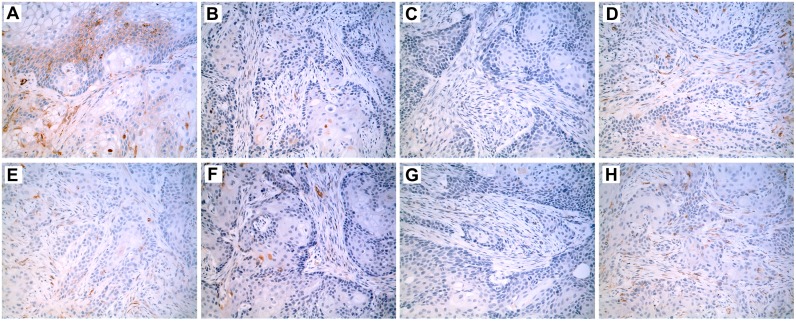
Fig 3. Immunohistochemistry of SPARC localization in tumor transplants generated from SPARC-transfected cell lines.
Immunohistochemical stain (brown) shows the location of SPARC expression within the tumor transplant tissues generated from the following UROtsa cell lines: (A) As#3-SPARC; (B) As#3-DEST; (C) As#6-SPARC; (D) As#6-DEST; (E) Cd#1-SPARC; (F) Cd#1-DEST; (G) Cd#4-SPARC; and (H) Cd#4-DEST cell lines. All images are shown with a 200X magnification.

