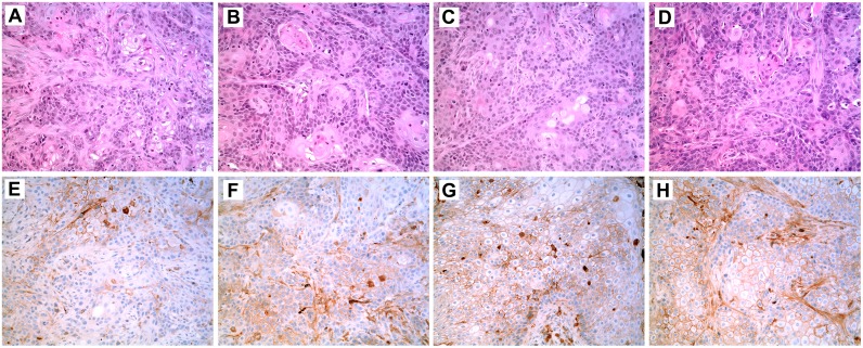
Fig 4. Histology and Immunohistochemistry of SPARC in Tumor Transplants Generated from Urospheres Isolated from As+3-and Cd+2-Transformed UROtsa Cell Lines and from As+3-and Cd+2-Transformed UROtsa Cell Lines Stably Transfected with SPARC.
Tumor transplants were generated from urospheres isolated from four transformed UROtsa cell lines: As#3 (A, E); As#3-SPARC (B, F); Cd#4 (C, G); and Cd#4-SPARC (D, H). The histology of the tumors generated from the urospheres is shown in (A-D) while the SPARC immunolocalization (brown stain) is shown in (E-H). All images are shown with a 200X magnification.

