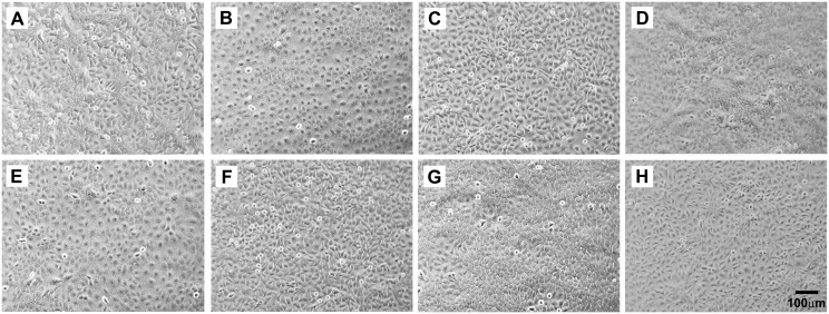
Fig 6. Phase contrast light microscopy of the SPARC-transfected and DEST-transfected UROtsa cell lines demonstrating epithelial morphology in all lines.
(A) As#3-SPARC; (B) As#3-DEST; (C) As#6-SPARC; (D) As#6-DEST; (E) Cd#1-SPARC (F) Cd#1-DEST; (G) Cd#4-SPARC; and (H) Cd#4-DEST. The magnification of all images corresponds to the bar shown in H (100 μm).

