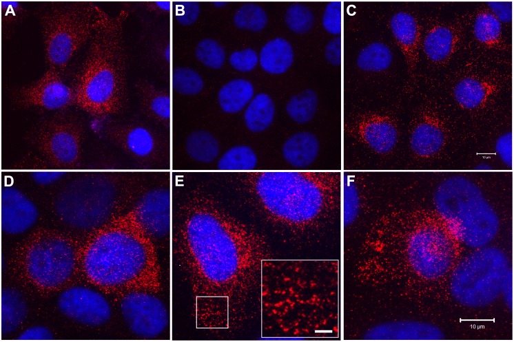
Fig 7. Intracellular localization of SPARC protein by immunofluorescent staining and confocal microscopy.
SPARC (red) immunostaining as well as DAPI (blue) staining to identify all cells in the field is shown for the following UROtsa cell lines: (A) Parent; (B) Cd#4-DEST (blank vector); and (C) Cd#4-SPARC cells. Higher magnification images are shown for: (D) As#3-SPARC, (E) As#6-SPARC, and (F) Cd#1-SPARC cells. The inset shown in (E) for the As#6-SPARC cells shows a high magnification image of the region of cytoplasm indicated by the white box. This staining pattern is consistent with intracellular vesicles containing SPARC protein. Images in A-C correspond to bar in C (10μm), images in D-F correspond to the bar in F (10μm), and the bar shown in the inset corresponds to 2 μm.

