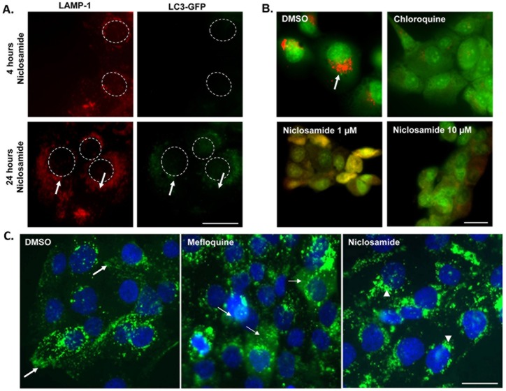
Fig 5. Lysosomes in niclosamide-treated cells are not leaky and are not autophagosomes.
(A) LC3-mCherry-GFP transfected DU145 cells were treated with 1 μM niclosamide for 4 hours or 24 hours and cells were fixed and stained for LAMP-1. Dashed circles represent nuclei. Arrows indicate increased colocalization of LC3-GFP with LAMP-1. Scale bars: 10 μm. (B) DU145 cells were incubated with Acridine Orange, washed, and treated with DMSO, niclosamide or chloroquine for 16 hours. Red represents acridine orange accumulation in the acidic lysosomes, as seen in DMSO condition. In cells treated with chloroquine or different concentrations of niclosamide, the leaked acridine orange in cytosol is no more concentrated in the acidic lysosomal compartment and therefore its color turns into green. Scale bars: 10 μm. (C) DU145 cells were loaded with dextran 40kDa (green) and then treated overnight with DMSO, 10 μM mefloquine, or 0.6 μM niclosamide. Cells were fixed and stained for DAPI (blue). In control cells lysosomes have a peripheral distribution (bold arrows), whereas in niclosamide treated cells lysosomes are intact and found near the nucleus (arrowheads). Mefloquine induces lysosomal membrane permeabilization, LMP, as manifested by green cytosolic haziness (thin arrows) and is the positive control. Scale bars: 10 μm

