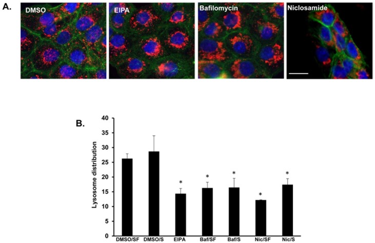
Fig 6. Bafilomycin A1, a specific inhibitor of vacuolar-type proton pump, induced perinuclear distribution of lysosomes.
(A) DU145 cells were treated for 16 hours with DMSO, 25 μM EIPA, 0.1 μM bafilomycin, 1 μM niclosamide or 50 μM chloroquine. Then, pH was dropped to 6.4 in all conditions for an additional 2 hours. Cells were then fixed and immunostained for LAMP1 (red), DAPI (blue) and actin (green). Scale bars: 10 μm. (B) DU145 cells were treated for 16 hours with DMSO, bafilomycin A1 (Baf), niclosamide (Nic), or EIPA in serum free (SF) and complete media (S). Lysosome distribution was calculated “mean ring spot count channel 3” using the Cellomics Imager. Error bars represent the SD from at least 3 independent experiments. * denotes statistical significance (p<0.05) relative to treatment with DMSO.

