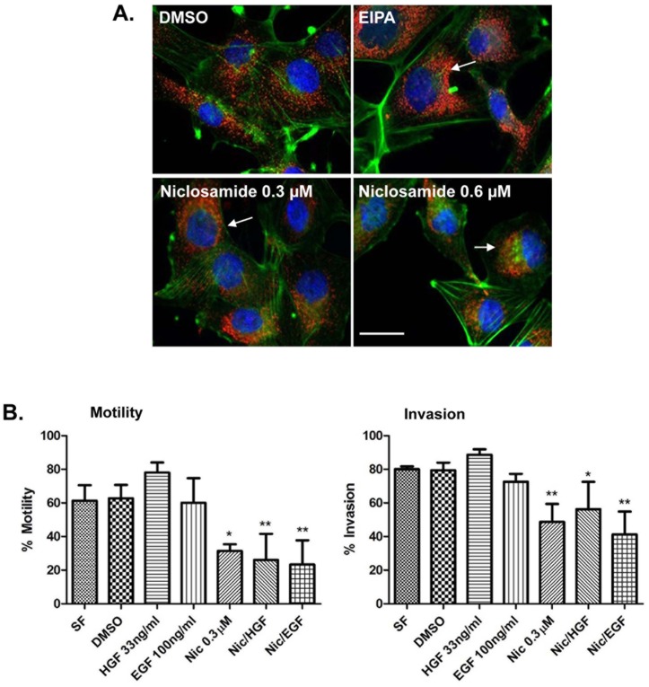
Fig 7. Niclosamide inhibits lysosome trafficking, motility, and invasion in glioma A172 cells.
(A) A172 cells were treated for 8 hours with DMSO, 25 μM EIPA, or varying concentrations of niclosamide diluted in low pH media (pH 6.4). Next, cells were fixed and stained for LAMP-1 (red), actin (green) and DAPI (blue). Lysosomes in DMSO control have a peripheral location whereas in niclosamide or EIPA-treated cells they are located around the nucleus (as indicated by arrows). Scale bars: 10 μm. (B) A172 cells were plated in collagen-coated 96 well plates and allowed to form a confluent monolayer prior to wounding. Next, 20% matrigel was added to the wells for which invasion is to be studied. Cells were allowed to migrate or invade in the presence of DMSO, 33 ng/mL HGF or 100 ng/mL EGF in the presence or absence of 0.3 μM niclosamide. Motility and invasion were calculated by IncuCyte Imager and the relative wound density percentage at 24 hours post wound is shown. Error bars represent the SD from at least 3 independent experiments. * p<0.05 treatment versus serum free or DMSO, ** p<0.01 treatment versus serum free or DMSO.

