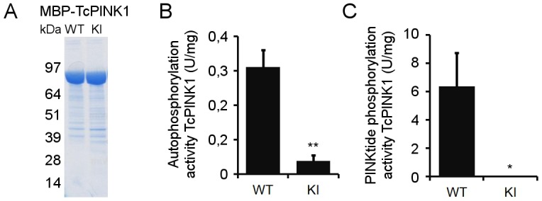
Fig 2. TcPINK1 autophosphorylates and phosphorylates PINKtide.

(A) Purity of E. coli-expressed WT and KI TcPINK1 was evaluated by Coomassie staining. Both forms of TcPINK1 are equally enriched. (B) Quantification of [γ-32P]-ATP in vitro autophosphorylation of purified WT or KI TcPINK1. (C) Quantification of [γ-32P]-ATP in vitro phosphorylation of PINKtide by purified WT or KI TcPINK1 (mean ± SEM, n = 4 independent experiments). Statistical significance was calculated between WT and KI TcPINK1 using Student’s t-test (*: p-value < 0.05; **: p-value < 0.01).
