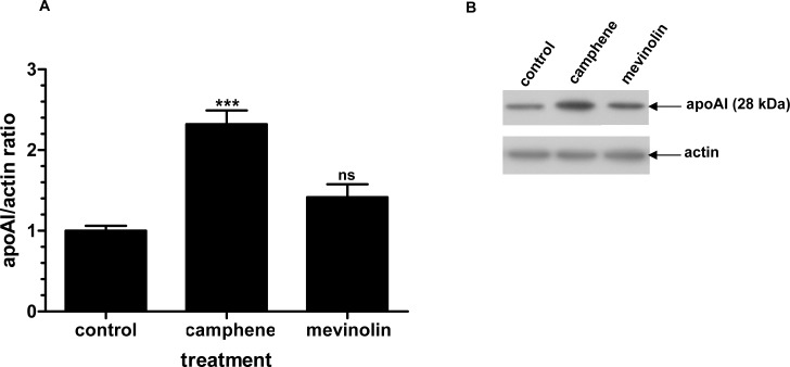
Fig 8. Effect of camphene and mevinolin on the protein expression of apoAI.
Panel A. On day 7, HepG2 cells were incubated for 48 h with camphene (37 μM) and mevinolin (37 μM) in DMEM containing 10% LPDS. Total cell protein was extracted and 50 μg of protein were separated by SDS PAGE and analyzed by Western immunoblotting using goat anti-apoAI antibody as described. Mouse β-actin was used to control for equal loading and normalization. Relative intensity of the bands was quantified using Phosphorimager. Values express the apoAI/actin ratio and are calculated by comparison to control samples the value of which is defined as 1.Values are means ± SD of three independent experiments in triplicates. p<0.001 (***) and ns (non significant) vs control by the Student-Newmann-Keuls test. Panel B. A representative Western blot is shown.

