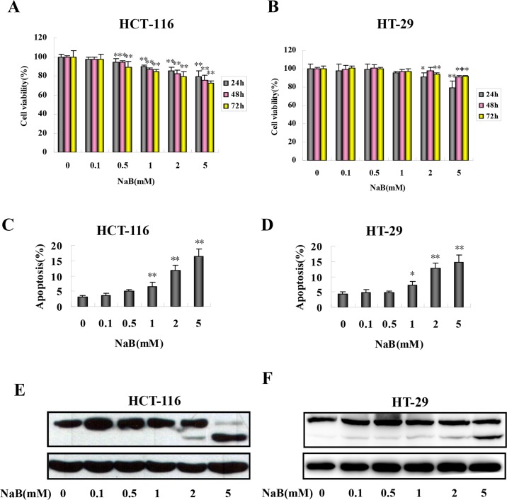
Fig 1. Sodium butyrate inhibited proliferation and induced apoptosis in colorectal cancer cells.
HCT-116 (A) and HT-29 (B) cells were treated with the indicated concentrations of sodium butyrate (NaB) for 24 (grey bars), 48 (pink bars), or 72 (yellow bars) hours and cell proliferation was assessed. HCT-116 (C, E) and HT-29 (D, F) cells were treated with the indicated concentrations of NaB for 24 h. C.D. The percentage of apoptotic cells in HCT-116 (C) and HT-29 (D) cells was quantified in three independent experiments using an annexinV/PI assay (annexinV+/PI-) by flow cytometry and expressed as mean ± SD. One-way ANOVA was used to compare between the control cells and NaB treatments. *p<0.05, ** p<0.01 compared to control. E.F. Representative Western blots of full-length and cleaved PARP products in HCT-116 (E) and HT-29 (F) cells.

