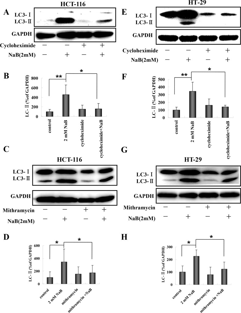
Fig 8. Cyclohexamide and mithramycin blocked sodium butyrate induced autophagy in colorectal cancer cells.
HCT-116 or HT-29 cells were treated with 10 μg/mL cycloheximide or 0.1μM mithramycin for 30 min and then with 2mM sodium butyrate (NaB) for 24 h. Representative Western blots of the expression of LC3-II are shown. The level of LC3-II expression was quantified by densitometry and normalized to GAPDH (ratio of LC3-II:GAPDH). The fold change from control cells is shown. Means and standard deviation (SD) of three independent experiments are shown. One-way ANOVA was used for statistical analysis. * P<0.05, ** p<0.01, compared to the control group. (A-B) HCT-116 cells treated with cyclohexamide; (C-D) HCT-116 cells treated with mithramycin; (E-F) HT-29 cells treated with cyclohexamide; (G-H) HT-29 cells treated with mithramycin.

