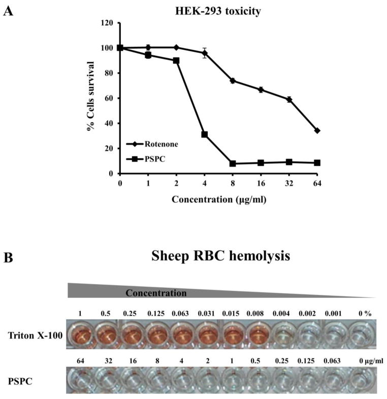
Figure 4. Cytotoxicity and hemolytic activity of PSPC.
(A) The viability of HEK-293 cells was measured after treatment with serially diluted concentrations (1–64 μg/ml) of PSPC or the mitochondrial toxin rotenone (positive control). Cell viability was measured spectrophotometrically by detecting degradation of WST-1 dye into formazan by viable cells, which produces measurable color.
(B) Sheep erythrocytes were treated with serial dilutions of Triton X-100 (.001–1%) or PSPC (0.063–64 μg/ml).

