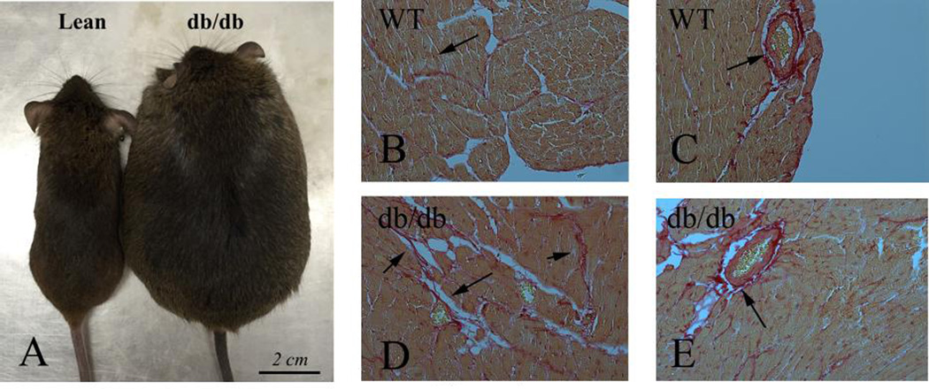
Figure 1.
Cardiac fibrosis in experimental models of diabetes. A. db/db mice develop severe obesity and diabetes, associated with myocardial fibrosis. B–E. Sirius red staining labels collagen (red – arrows) in the cardiac interstitium (B) and in perivascular areas (C) in lean and db/db mice (D–E). db/db animals exhibit expansion of the interstitial space (D) and perivascular accumulation of collagen (E).

