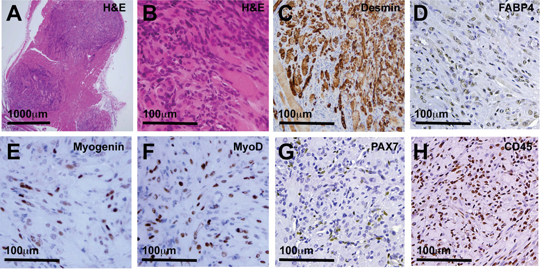
Figure 1.
Microscopic tumor foci in R26-SmoM2(+/−);CAGGS-CreER mouse skeletal muscle contain phenotypically heterogeneous cells. (A) Random muscle sections obtained from obtained from R26-SmoM2(+/−);CAGGS-CreER mice contained numerous microscopic tumor foci. (B) Tumors contained large, multinucleated cells with abundant cytoplasm interspersed with small round blue cells. (C) Desmin-expressing, (D) FABP4-expressing, (E) Myogenin-expressing, (F) MyoD-expressing, (G) PAX7-expressing, (H) CD45-expressing tumor cells were interspersed with many Desmin-, FABP4-, Myogenin-, MyoD-, Pax7- and CD45-negative tumor cells. (C) Desmin expression in tumors often localized to larger cells with abundant cytoplasm. Tumors contained numerous cells expressing (D) adipocytic cell lineage markers (FABP4) and (H) hematopoietic-lineage markers (CD45).

