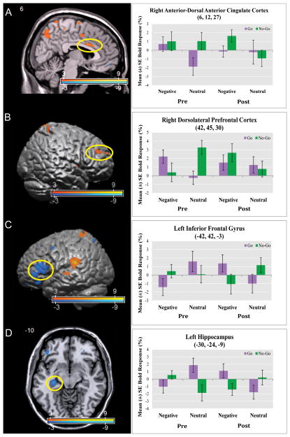
Figure 1. Increased Dorsal Anterior Cingulate and Dorsolateral Prefrontal Cortex Activation and Decreased Inferior Frontal Gyrus and Hippocampus Activation during Behavioral Inhibition in the Context of Negative Emotional Processing Post- vs. Pre-Transference-Focused Psychotherapy.
Panels A–D depict the interaction [(post-treatment vs. pre-treatment) × (negative vs. neutral) × (no-go vs. go)] (Supplementary Table 2 and 3). Statistical parametric maps are thresholded at a voxelwise p-value of 0.01. Following treatment with Transference Focused Psychotherapy (TFP), borderline personality disorder patients demonstrated relative increased activation in the (Panel A) right anterior-dorsal anterior cingulate cortex (voxel-wise p-value=0.001; corrected p-value=0.022) and the (Panel B) right dorsolateral prefrontal cortex (voxel-wise p-value=0.001); relative activation decreases following treatment were noted in the (Panel C) left inferior frontal gyrus (voxel-wise p-value < 0.001) and the (Panel D) left hippocampus (voxel-wise p-value = 0.001).

