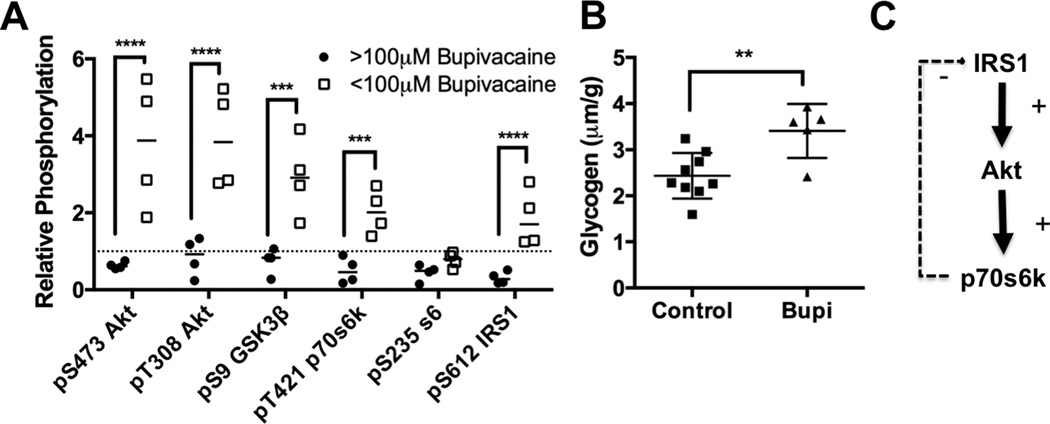
Figure 3. Recovery dependent cardiac protein phosphorylation.
A. Densitometry of phospho-proteins in insulinergic pathway in recovered animals with <100μM cardiac bupivacaine (n=4) compared to densitometry during toxicity (>100μM bupivacaine, n=4); ***p<0.001, ****p<0.0001, Sidak post-hoc test. Proteins blotted for include protein kinase B (Akt) phosphorylated at S473, Akt phosphorylated and T308, total Akt, glycogen synthase kinase alpha (GSK-3α) and beta (GSK-3β) phosphorylated at S21 and S9 respectively, p70 s6 kinase (p70s6k) phosphorylated at T421, ribosomal protein s6 (s6) phosphorylated at S235, insulin receptor substrate 1 (IRS1) phosphorylated at S612, and total glyceraldehyde-3-phosphate dehydrogenase (GAPDH) as loading control. B. Cardiac glycogen concentration for control hearts (n=9) and bupivacaine treated hearts in recovery (n=5) **:p=0.01 by two-sided t-test. C. Schematic of feedback pathway underlying sensitization of insulinergic signaling demonstrating how loss of feedback from p70s6k (and other kinases) to IRS1 leads to a sensitization and amplified activation of downstream targets including Akt.

