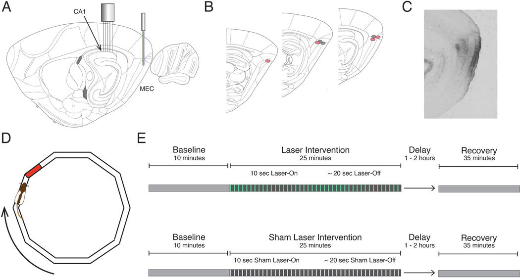
Figure 1. Schematic of the inactivation procedure and task design.
A, Optogenetic inactivation of the dorsocaudal medial entorhinal cortex (MEC) was achieved using a 532 nm laser to activate the inhibitory opsin ArchT. Simultaneous electrophysiological recordings were performed in the ipsilateral dorsal CA1. B, Reconstruction of optic fiber tip locations. Red and gray circles correspond to rats expressing ArchT or GFP, respectively. C, Representative sagittal section from a rat expressing ArchT, indicating the spread of viral infection in the MEC. D, For every phase of the experiment, rats completed laps on an elliptical track for a food reward in a reliably rewarded location (red). E, Experimental paradigms for both Laser (top) and Sham Laser (bottom) intervention days consisted of a Baseline period, an Intervention period (constituted by 30 alternating 10-second Laser-On and ~20-second Laser-Off intervals), and a Recovery period.

