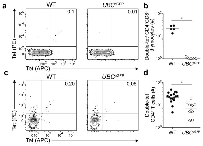
Figure 6. Expression of a self-epitope by thymic antigen-presenting cells induces intrathymic clonal deletion.
(a) Contour plots of tet-PE versus tet-APC staining of tetramer-enriched CD4+CD8− thymocytes from WT or UBCeGFP mice.
(b) Number of CD4+CD8− double-tet+ cells in the thymuses of WT or UBCeGFP mice. Horizontal bars indicate geometric mean values and circles represent individual mice. Data were pooled from two independent experiments, n=5 total mice per group. (* P < 0.0001 by unpaired t-test of log10 transformed data).
(c) Contour plots of tet-PE versus tet-APC staining of tetramer-enriched CD4+ T cells from pooled spleens and lymph nodes of WT or UBCeGFP mice.
(d) Number of CD4+ double-tet+ cells in pooled spleens and lymph nodes of WT or UBCeGFP mice. Horizontal bars indicate geometric mean values and circles represent individual mice. Data were pooled from 4 independent experiments, n=10–17 total mice per group. Data for WT mice are replotted from Figure 2b. (* P < 0.0001 by unpaired t-test of log10 transformed data).
Numbers on each plot in (a) and (c) indicate the percent of double-tet+ cells.

