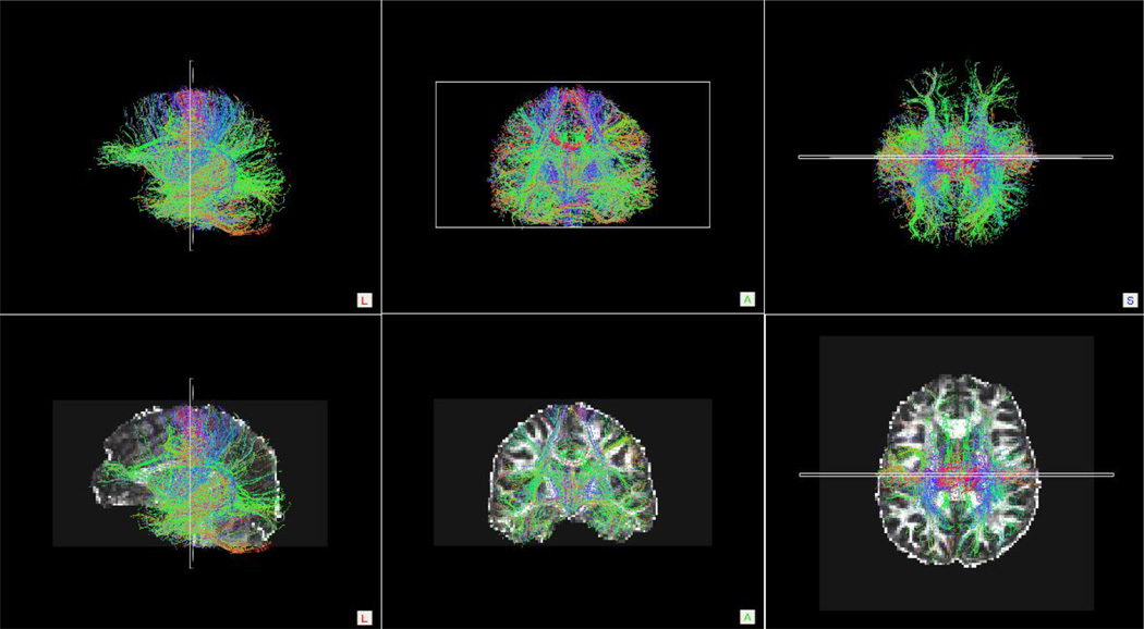
Figure 6.
Whole Brain Streamline Tractography Results
Top row: Sagittal, coronal and axial orientation images of whole brain streamline tractography conducted on a single control participant. Bottom row: To provide anatomical references, the images from the top row were overlaid onto the participant’s FA-weighted diffusion image.

