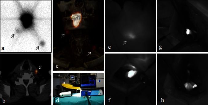
Fig. 1.
Preoperative and intraoperative SN imaging. LSG (a), axial SPECT/CT (b), and the 3-D reconstructed SPECT/CT (c) in a patient with a well-lateralized tumor on the right anterior tongue showing tracer drainage to an ipsilateral SN in level 1 and directly to a contralateral SN in level 3 (arrows). The latter SN in level 3 in the left neck side was visible transcutaneously (e). The Fluobeam 800 NIR camera (d) designed for ICG imaging. The NIR camera entered the surgical field in a sterile cover, and real-time video imaging was presented for the surgical team on a clinical screen. When using the NIR camera intraoperatively, the direct surgical light was turned off to improve the quality of the imaging. The system has a 750-nm excitation laser and LED white light illumination of the surgical field that does not inflict on the NIRF imaging. The hand-held camera head is maneuverable in all angles and has a ×10 zoom function. The autofocus function allows for flexible working distance. Intraoperative NIRF-guided identification and resection of SN (f, g, h)

