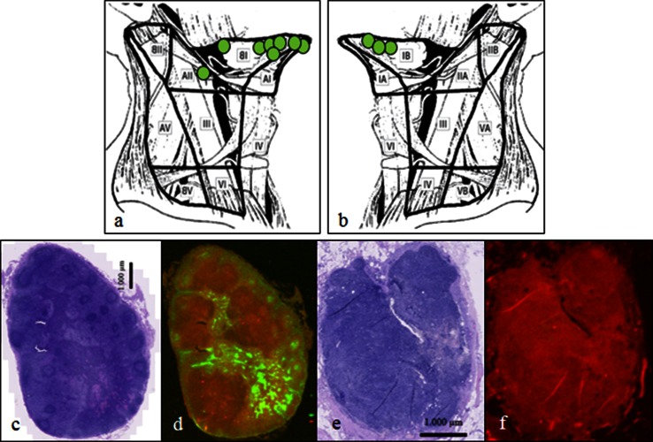
Fig. 2.
Intraoperative SN identification by NIRF imaging only. The exact anatomical location and neck level location in the ipsilateral (a) and contralateral (b) neck side of the 11 additional SNs identified only by fluorescence. H&E staining and fluorescence microimaging of a tissue section from a SN (c and d) and a non-SN (e and f) located intimately within the same cluster of lymph nodes in level 2a. None of the lymph nodes contains metastatic tumor. In the SN the microantomical distribution of the fluorescent tracer draining from the marginal sinus towards the medullary sinus is visualized. The non-SN is without any signal from ICG on NIRF microimaging

