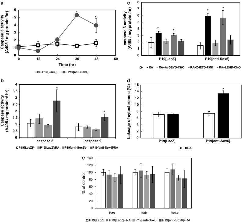
Fig. 2.

Sox6 suppression induced caspase 3 and 9 activation and leakage of cytochrome c. a Cells were incubated with 500 nM RA for 0–48 h, after which caspase 3-like activity was measured for 2 h using Ac-DEVD-pNA as the substrate. b Cells were cultured in bacterial-grade dishes with or without 500 nM RA for 36 h. Caspase 8- and 9-like activities were separately measured for 2 h using Ac-IETD-pNA as the substrate for caspase 8 and Ac-LEHD-pNA for caspase 9. c Cells were cultured in bacterial-grade dishes with or without 500 nM RA and the caspase inhibitors for 36 h, after which caspase 3-like activity was measured for 2 h using Ac-DEVD-pNA as the substrate. d Cells were cultured in bacterial-grade dishes with or without 500 nM RA for 36 h. Cytochrome c was measured by ELISA after subcellular fractionation. e Cells were cultured in bacterial-grade dishes with or without 500 nM RA for 36 h. The cytoplasmic Bcl-2 families were assessed by ELISA after subcellular fractionation. Asterisk indicates differences that were considered to be significant at P < 0.05 by Scheffe’s F test
