Figure 3.
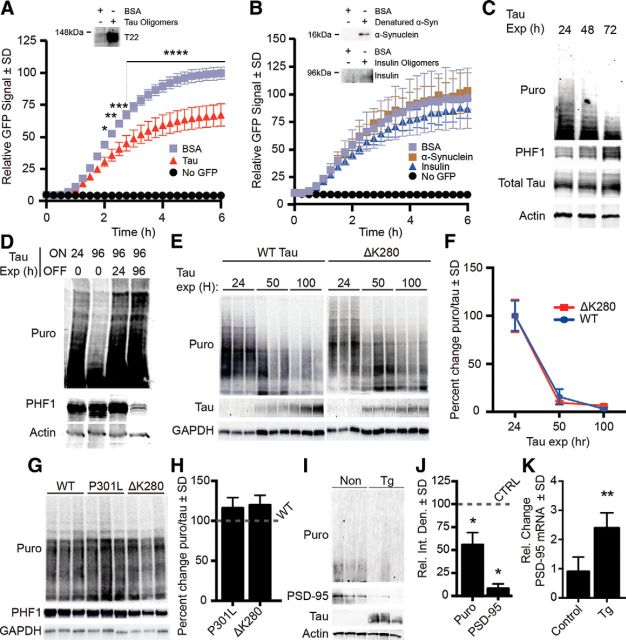
Pathological tau species decrease translation. A, IVT graph showing 32% reduction of translational output (after 6 h) in wells with tau oligomers compared with BSA control. B, Oligomeric α-synuclein and insulin did not affect translation. Immunoblots confirm the presence of oligomers in the translation assay; α-synuclein samples were denatured to yield monomers because high molecular weight oligomeric conformers are not detectable with anti-α-synuclein antibodies. C, Total and PHF1 tau are inversely proportional to the rate of protein synthesis. iHEK-Tau cells were stimulated with tetracycline to express tau for 4 d. Nascent proteins were tagged with puromycin. D, Cessation of tau expression rescues protein synthesis. Tau expression was induced with tetracycline for 24 or 96 h (lanes 1 and 2). At 96 h, puromycin levels decreased, whereas PHF1 increased. After continuous tau expression for 96 h (lanes 3 and 4), cells were washed and incubated with media lacking tetracycline, thereby halting tau expression for 24 or 96 h (lanes 3 and 4, respectively). E, Representative immunoblots showing the effect of wild-type (WT) and ΔK280 tau on protein synthesis. Changes in tau levels inversely correlated with puromycin. F, Quantification of E showing no significant difference in levels of protein synthesis between wild-type and ΔK280 mutant tau-expressing cells. G, SUnSET comparing the effect of P301L, ΔK280 tau, and WT in transiently transfected HEK293 cells. H, Quantification of G showing no significant difference between translation levels. I, Blot showing that overall translation (Puro) and PSD-95 are decreased in rTg4510 primary neurons. J, Quantification of I. Puromycin and PSD-95 were significantly decreased in rTg4510 neurons by 43 and 92%, respectively (*p < 0.05). K, Quantification of PSD-95 mRNA expression from rTg4510 neurons as measured by RT-PCR (**p = 0.0013).

