Figure 7.
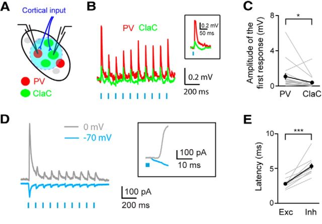
Cortical input evokes feedforward inhibition in ClaC neurons. A, Experimental configuration showing retrogradely labeled ClaC (green) neurons and genetically labeled PV (red) claustral neurons. ChR2 (blue) is expressed in corticoclaustral axons. B, An example of a paired recording from a ClaC neuron (green trace) and a PV neuron (red trace) during optogenetic activation of corticoclaustral afferents. Inset, Response to the first light flash shown at an expanded time scale. C, Summary data showing the amplitude of the first response in paired recordings of one ClaC neuron and PV neuron following optogenetic activation of corticoclaustral axons (n = 17, p = 0.0127258, sign test). On average, the postsynaptic potentials measured were larger in PV neurons than in ClaC neurons following optogenetic activation of corticoclaustral afferents. D, Voltage-clamp recording of a ClaC neuron held at −70 mV and at 0 mV during optogenetic activation of corticoclaustral afferents. Inset, Response to the first light flash of the train shown at an expanded time scale. Note the delayed onset of the outward current. E, The latency of the response onset for the inward component measured at −70 mV and the outward component measured at 0 mV. The latency was significantly longer for the outward component (inward: 2.79 ± 0.21 ms; outward: 5.33 ± 0.41 ms, n = 10 cells, p = 0.0003, paired t test).

