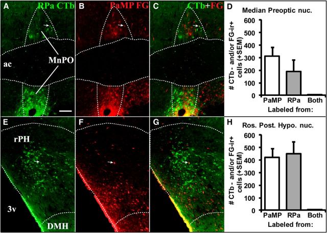
Figure 2.
Retrograde tracer labeling in MnPO (0.1 mm posterior to bregma; A–C) and rPH (3.3 mm posterior to bregma; E–G) following tracer injections in RPa (A, E, green-labeled cells) and PaMP (B, F; red-labeled cells) in Case 62. Superimposed photomicrographs of retrograde labeling from the two target regions (C, G) indicate very few cells displaying colocalization of the two distinct tracers in the same cells (white arrows). Counts of retrogradely labeled cells (+1 SEM) immunoreactive for CTb and/or FG in the MnPO (D) and rPH (H) indicated relatively similar cell numbers originating from the RPa and PaMP tracer injections, but very few cells colocalized the two tracers (Both). 3v, Third ventricle; ac, anterior commissure; DMH, dorsomedial hypothalamic nucleus. Scale bar (in A): A–H, 100 μm.

