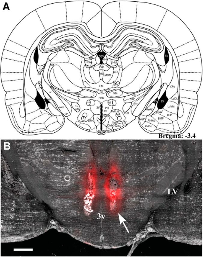
Figure 7.

A, Plate from Paxinos and Watson's rat brain atlas (Paxinos and Watson, 2004; their Fig. 62) representing the location of injector cannulae tips for ACSF (Veh) or muscimol injections in the repeated loud noise and restraint stress studies. B, Coronal section of a fresh-frozen brain slice (35 μm) following bilateral injections of 200 nl of a BODIPY TMR-X muscimol conjugate (red) targeting the rostral posterior hypothalamic nucleus (3.4 mm posterior to bregma). Note the restricted lateral dispersion of muscimol from the injector tips (white arrow), which appears more extensive in the ventrodorsal plane. 3v, Third ventricle; LV, lateral ventricle. Scale bar, 1000 μm.
