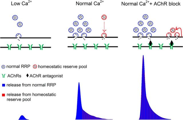
Figure 6.
Blocking postsynaptic AChRs mobilizes a homeostatic reserve pool of vesicles at the mouse NMJ. Illustration of single action potential evoked synaptic vesicle release (top diagram) and its corresponding release profile obtained by deconvolution of evoked current (bottom). When extracellular Ca2+ entry is low, the only pool of vesicles participating in release is the synchronously released pool of vesicles (blue vesicles). Both release probability and the number of releasable vesicles are low. With the elevation of extracellular Ca2+ level, there is an increase in the pool of synchronously released vesicles. The time course of release of these vesicles is indicated by vertical blue lines in the deconvolution profile. In normal Ca2+ levels, there is also recruitment of the homeostatic reserve pool (red vesicles), but most vesicles in this pool are released slowly, after the peak of vesicle release is over (red dotted arrow in diagram, vertical red lines in deconvolution profile). Following partial block of AChRs, the homeostatic reserve pool becomes primed and is released as rapidly as the synchronously released pool (superimposed red and blue vertical lines, deconvolution profile). The homeostatic reserve pool is small and must be continuously replenished through vesicle recycling (red arrow).

