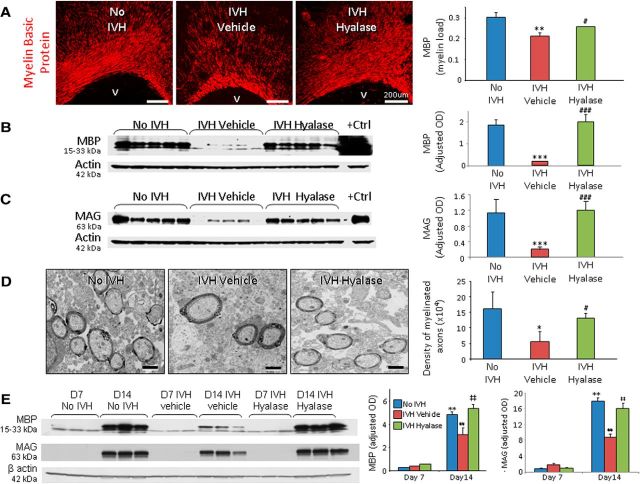
Figure 6.
Hyaluronidase treatment restores myelination in rabbits with IVH. A, Representative immunofluorescence of MBP in the corona radiata of day 14 pups. Volume fractions of MBP were higher in the corpus callosum and corona radiata of hyaluronidase-treated pups compared with vehicle-treated controls with IVH. V, Ventricular side. B, Typical Western blot analysis for MBP in the forebrain of premature rabbit pups, as indicated, at day 14. Adult rat brain was used as a positive control. Each lane represents a lysate from a whole coronal slice taken at the level of midseptal nucleus of one brain. MBP expression was higher in hyaluronidase-treated pups compared with vehicle-treated pups. C, Western blot analysis for MAG in the forebrain of pups as indicated at day 14. Adult rat brain was used as a positive control. MAG expression was higher in hyaluronidase-treated pups compared with vehicle-treated controls. D, Typical electron micrograph from rabbit pups without and with IVH, and pups with IVH treated with hyaluronidase at day 14. Note that myelinated axons were fewer in pups with IVH compared to controls without IVH and that hyaluronidase treatment significantly increased the number of myelinated axons in pups with IVH. E, Representative Western blot analyses for MBP and MAG for three groups of pups (as indicated) at days 7 and 14. Note the similar expression of MBP and MAG in the three sets of pups at day 7. MBP levels at day 14 were ∼10-fold higher compared to day 7 in pups without IVH and hyaluronidase-treated pups with IVH. *p < 0.05 (pups with vs without IVH), **p < 0.01 (no IVH pups day 7 vs day 14), ***p < 0.001 (pups with vs without IVH); #p < 0.05 (vehicle- vs hyaluronidase-treated pups with IVH), ##p < 0.001 (pups with IVH day 7 vs day 14), ###p < 0.001 (vehicle- vs hyaluronidase-treated pups with IVH); ‡‡p < 0.001 (pups with IVH day 7 vs day 14). Scale bars: A, 200 μm; D, 1 μm. Data are mean ± SEM (n = 5 each group).

