Figure 3.
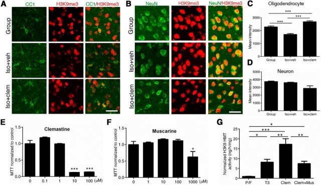
Clemastine increases levels of repressive histone modification H3K9me3 in oligodendrocytes. A, B, Confocal images of H3K9me3 (red) and CC1+ (green) oligodendrocytes (A) or NeuN+ (green) neurons (B) in PFC of group-housed (Group), iso+veh, and iso+clem mice. Scale bar, 25 μm. C, Quantifications of H3K9me3 immunoreactivity in CC1+ oligodendrocytes in the PFC [n = 167 cells from 2 group-housed (Group) mice, n = 143 cells from 3 iso+veh mice, and n = 258 cells from 3 iso+clem mice]. ***p < 0.001 by one-way ANOVA followed by Tukey's post hoc test. D, Quantifications of H3K9me3 immunoreactivity in NeuN+ neurons in the PFC (n = 293 cells from 2 group-housed (Group) mice, n = 436 cells from 3 iso+veh mice, n = 411 cells from 3 iso+clem mice). E, F, The dose-dependent toxicity of clemastine (E) and muscarine (F) was assessed in primary mouse oligodendrocyte cultures, using MTT assays. G, H3K9 methyltransferase activity increased when OPCs were treated for 72 h with thyroid hormone (T3, 45 nm) or clemastine (1 μm) compared with mitogens PDGF A chain homodimer and bFGF (P/F). The increase of H3K9 methyltransferase activity by clemastine was attenuated when OPCs were cotreated with clemastine (1 μm) and muscarine (100 μm). Fold changes were calculated after normalizing to activity in proliferating condition (P/F). n = 3 separate cultures per condition. *p < 0.05, **p < 0.005, ***p < 0.001 by one-way ANOVA followed by Tukey's post hoc test. Data are mean ± SEM.

