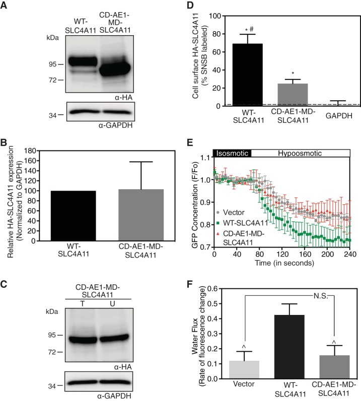
Fig. 5.
Fusion to AE1 CD restores MD-SLC4A11 localization but not function. HEK-293 cells were transfected with cDNA encoding HA epitope-tagged WT-SLC4A11 and CD-AE1-MD-SLC4A11 chimera (AE1 residues 1–354 fused to SLC4A11 residues 341–891). A: cell lysates (50 μg of protein) were immunoblotted with anti-HA (α-HA) and anti-GAPDH (α-GAPDH) antibodies. B: densitometry of expression, normalized for GAPDH abundance and standardized to WT-SLC4A11. C: transfected cells were labeled with membrane-impermeant sulfo-NHS-SS-biotin for cell surface biotinylation assays, and cell lysates were probed on immunoblots. D: fraction of WT-SLC4A11, CD-AE1-MD-SLC4A11, and GAPDH labeled by sulfo-NHS-SS-biotin. Values are means ± SE (n = 3). Dashed line indicates level of biotinylated GAPDH, which represents the background level for the assay. E: HEK-293 cells, grown on coverslips, were transiently cotransfected with cDNA encoding GFP and vector or SLC4A11 chimera. Cells were perfused with isosmotic and hyposmotic media. F, fluorescence. F: rate of fluorescence change, which represents water flux. Values are means ± SE from 3 independent experiments with 8–10 cells measured per coverslip. *P < 0.05 vs. GAPDH. #P < 0.05 vs. CD-AE1-MD-SLC4A11. P̂ < 0.05 vs. WT-SLC4A11. NS, not significant (P > 0.05).

