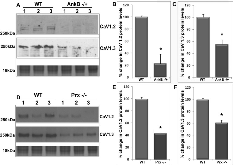
Fig. 8.
Decreased levels of L-type Ca2+ channel proteins in AnkB−/+ and Prx−/− mouse lenses. A–C: significantly decreased voltage-gated calcium channels CaV1.2 (A and B) and CaV1.3 (A and C) protein content in AnkB−/+ lenses (membrane-enriched fraction from 5-mo-old mice) compared with WT lenses based on immunoblot quantification. D–F: significantly decreased CaV1.2 (D and E) and CaV1.3 (D and F) channel protein content in Prx−/− lenses (5 mo old) compared with WT lenses. In A and D, lanes 1–3 show representative immunoblot profiles from three independent specimens. Staining of proteins from membrane-enriched fraction (in the range of 16–18 kDa) was used as a loading control. Values in B, C, E, and F are means ± SE of 4–6 independent observations. *P < 0.05.

