Fig. 2.
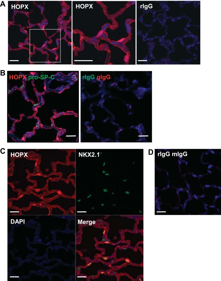
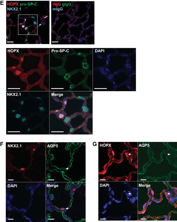
Immunofluorescence for alveolar type 1 (AT1) and 2 (AT2) cell markers in normal rat lung. A: left, immunofluorescence for homeodomain only protein x (HOPX, red) shows both nuclear and cytoplasm/membrane staining. Middle, magnified views of the rectangles shown on left. Right, negative control is rIgG. DAPI (blue) is the nuclear counterstain. Bar = 20 μm. B: double immunofluorescence shows HOPX (red) does not colocalize with pro-SP-C (green). Bar = 20 μm. C: double immunofluorescence shows nuclear HOPX (red) colocalizing with NKX2.1 (green). Bar = 20 μm. D: negative controls for C are rIgG and mIgG. E: triple immunofluorescence shows that NKX2.1 (light blue) colocalizes with either nuclear HOPX (red) or pro-SP-C (green), but not both. High magnification of rectangle is shown on bottom with individual channels. Bar = 20 μm. F: immunofluorescent localization of NKX2.1 (red) and AQP5 (green). Bar = 20 μm. Some cells are seen that colocalize both markers (arrows). G: immunofluorescent localization of HOPX (red) and AQP5 (green). Bar = 20 μm. Although most cells coexpress both markers, some do not (arrows).


