Fig. 4.
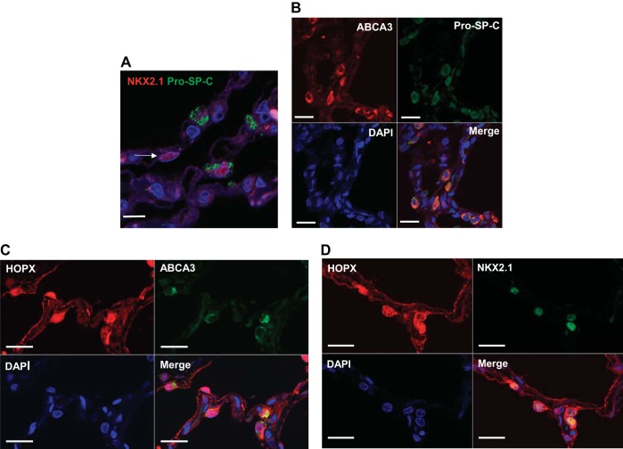
AT1 and AT2 cell markers in human lung tissue. A: not all NKX2.1+ cells colocalize with pro-SP-C in normal human lung (arrow, confocal z-stack image). DAPI (blue) was used to identify nuclei. Bar = 20 μm. B: ABCA3+ cells (>95%) coexpress pro-SP-C. Bar = 20 μm. C: many cells that express HOPX (red) also express the AT2 cell marker ABCA3 (green). Bar = 20 μm. D: most cells that express nuclear HOPX are also NKX2.1+. Bar = 20 μm.

