Fig. 1.
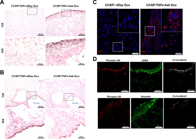
Phosphorylated p70 ribosomal S6 kinase (S6K) expression in the lung following transforming growth factor (TGF)-α overexpression. Clara cell secretory protein (CCSP)/TGF-α mice were administered doxycycline (Dox) to induce TGF-α expression. Immunostaining for phosphorylated S6 was performed on the lung sections of TGF-α mice administered Dox for 4 days or 4 wk comparing the subpleural compartment (A) with the airway/adventitial area (B). Insets demonstrate higher magnification of cells shown on bottom. C: immunofluorescence for phosphorylated S6 (red) on the 4-wk sections in the parenchyma of CCSP/− controls and CCSP/TGF-α mice. Insets demonstrate higher magnification of cells along the subpleural surface. D: immunofluorescence for phosphorylated S6 (red), α-smooth muscle actin (SMA), or vimentin (green) and colocalization (white) on the subpleural fibrotic regions for CCSP/TGF-α mice following 4 wk of Dox reveal colocalization of pS6 with mesenchymal cells. Staining is representative of individual sections from 4–8 mice/group.

