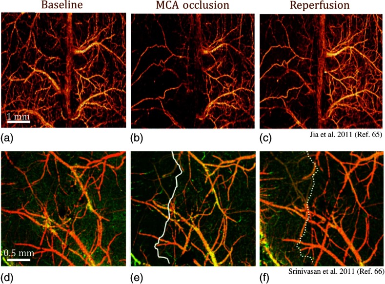
Fig. 6.
Volumetric OCT angiography imaging of the cortex during ischemic stroke. En face MIP of UHS-OMAG images (a) during baseline, (b) progressive focal ischemia developed during MCAO, and (c) 30 min after onset of reperfusion.65 En face MIP of OCT angiograms (d) at baseline, (e) during MCAO, and (f) 60 min after reperfusion. During MCAO (e), a capillary nonperfused region is apparent, as marked with a solid white line.66

