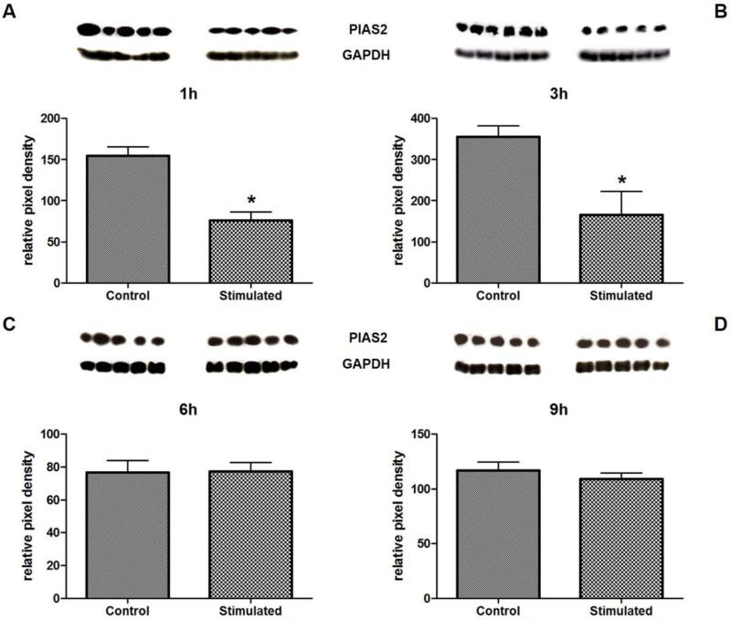
Figure 1.
Verification of antibody microarray data for PIAS2 by Western blot. Western blots were performed with lysates from HUVEC stimulated for 1h (A), 3h (B), 6h (C), and 9h (D) with 10−7M Ang-(1-7). Protein quantities were measured by densitometry and were normalized to GAPDH levels. Cells stimulated with the solvent were used as control.

