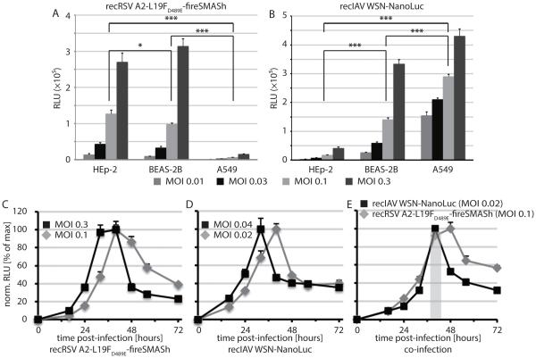
Figure 4.
Infection conditions for synchronized RSV and IAV reporter expression. A and B) Luciferase activities in three different human respiratory host cell lines 44 hours post-infection at the specified MOIs with recRSV-L19FD489E-fireSMASh (A) or recIAV WSN-NanoLuc (B; N = 4; means ± SD are shown). Two-way ANOVA with Tukey’s multiple comparison post-tests were carried out to assess statistical significance of sample divergence. Results are shown for MOI 0.1 (A) and 0.04 (B); *: p < 0.05; ***: p < 0.01. C-E) Reporter activity profiles after infection of BEAS-2B cells singly with recRSV-L19FD489E-fireSMASh (C) or recIAV WSN-NanoLuc (D), or after co-infection with both strains at an MOI of 0.1 (RSV) and 0.02 (IAV), respectively (E). Values represent cell-associated luciferase activities and were normalized to the highest signal of each series (N = 3; means ± SD are shown); grey shaded area in (D) marks the time window post-infection when signal intensities of both luciferase reporters are ≥80% of max.

